Showing posts with label congenital heart disease. Show all posts

2/27/2014

3D-Printed Heart Saves The Life Of A 14 Month Old Kid


3D-printing is one of the greatest technologies to come out in the past few years. It has been used by various industries in different fields and has allowed us to unveil new horizons. There is no doubt that this technology is revolutionizing the world, but now it’s even saving lives.


14-month old, Roland Lian Cung Bawi, from Owensboro, Kentucky was diagnosed with four congenital heart defects. He was admitted into the Kosair Children’s Hospital in Louisville, Kentucky where doctors knew immediately that the boy would need surgery. The only obstacle in the way was that doctor’s could not tell precisely what was wrong until they were in the middle of the surgery.


Surgeon Erie Austin realized that creating a 3D model of the boy’s heart would help study the defects and save the boy’s life. Dr. Austin called the Speed School of Engineering at the University of Louisville and told her of the problem and suggested a solution. The engineering department provided her a MakerBot 3D printer which would allow them to make a 3D model of the boy’s heart using 2D CT scans just as Dr. Austin had imagined. Tim Gornet, manager of the University’s Rapid Prototyping Center, was the one who the surgeon reached out to and his positive response helped save the boy’s life.
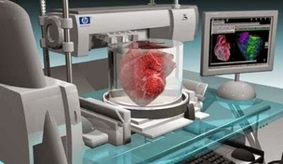

The Rapid Prototyping Center created a model made from a polymer known as ‘Ninja Flex’ which was 1.5 times larger than the boy’s heart (for easier inspection). Three general pieces of flexible filaments were made in the $2500 printer in 20 hours. The replica cost $600 to create, according to Gornet. The model helped doctors study the defects and come up with solutions before the critical surgery and on February 10th, 2014, Roland’s heart was repaired by Dr. Austin in what is the first use of 3-D printing for treating a pediatric heart patient.


The boy was released from the hospital on February 14th and and returned on February 21st for monitoring checks which showed good results. Thanks to Dr. Austin’s forward thinking and the savvy engineering team from the University of Louisville, a young life was saved and a new step was taken in medical technology that can save countless lives in the near future.


5/31/2013

Creating Valve Tissue Using 3-D Bioprinting

Aortic valve disease (AVD) is a serious health condition that affects people of all ages. Congenital heart valve defects are especially dangerous for newborns and can be fatal if left untreated. The most recommended treatment for AVD is surgical replacement of the defective valve. Although prosthetic valve replacement is the standard procedure for adults, these prosthetic devices are inadequate for younger adults and growing children. Tissue engineering has the potential to address these limitations of nonliving prosthetics (as well as human donor supply shortages) by providing living tissues that can grow, remodel, and integrate with the patient.
A critical requirement for tissue-engineered heart valves is that the engineered valve must be able to mimic the physiological function of the native valve, including the natural geometry and performance of the valve root, cusps, and sinus wall, all of which are essential for healthy coronary blood flow. Tissue-engineered heart valves must also have the same intrinsic asymmetry as the root, which prevents cusp deterioration.
A popular technique in the advanced manufacturing world, 3-D printing, has been modified to create precise, 3-D structures from living tissue. This 3-D "bioprinting" technology has been used by researchers at Cornell University to fabricate living heart valvesthat possess the same anatomical architecture as the original valve. This research is described in a paper by Jonathan T. Butcher.


3-D Construction

Biomaterials have been adapted for 3-D bioprinting, including co-polymer hydrogels. Alginate, for example, is a naturally occurring anionic polymer with many attractive features for biomedical applications, including low cost, excellent biocompatibility, low toxicity, and a variety of cross-linking capabilities. "Alginate-based hydrogels are particularly attractive for bioprinting because of their broad range of viscosities at room temperature," says Butcher. "This is important because hydrogels have tight requirements with regard to viscosity and gelling speed for accurate printing." Butcher's team conducted bioprinting that utilized an alginate/gelatin hydrogel system that included smooth muscle cells and valve interstitial cells.
A dual-syringe system was used to mimic the structure of the valve root and leaflets, two key valve structures. The team successfully fabricated living aortic valve conduits with strong anatomical resemblance to the native valve. The results demonstrate that anatomically complex, heterogeneously encapsulated aortic valve hydrogel conduits can be fabricated with 3-D bioprinting.

Moving Forward

"3-D tissue printing combines the disciplines of quantitative image analysis, computer-aided design, and manufacturing to develop a real entity—in this case, a living tissue—in a fraction of the time that any other traditional mechanical engineering process would," says Butcher. "Many people may spend their entire thesis working on just one part of this process, but without performing the whole process to completion, they won't know how to improve the system. Researchers gain a much greater appreciation for this systematic approach and can build a larger toolset, while having much more fun being creative in the process."
Butcher believes bioprinting will gain much more traction in the tissue engineering and biomedical community over the next five years, "potentially even becoming the standard in complex tissue fabrication," he adds. "I hope that people use this technology in the future to target a higher level of tissue complexity, like glandular and highly vascularized and innervated functional tissues."